For many years, fascia was considered little more than the connective tissue that wrapped around muscles, providing support and structure, but today, advances in anatomical and biomechanical research have transformed this view.
Rather than being an inert packing material, fascia is now recognised as a dynamic, living tissue that plays an essential role in movement, force transmission, proprioception (the body’s awareness of its position in space), and pain perception (Schleip et al., 2012; Stecco et al., 2018; Adstrum et al., 2017).
This growing understanding has led to increasing interest in myofascial therapies within both human and veterinary medicine. In horses, myofascial release (MFR) is becoming an increasingly recognised component of physiotherapy, used alongside rehabilitation exercises, therapeutic massage and other manual therapies to optimise movement, improve comfort and support performance.
Although scientific evidence in horses is still emerging, research suggests that the fascial system may play a far more active role in musculoskeletal health than previously believed (Harrison et al., 2015; Harrison and Elbrønd, 2018).
What is fascia?
Fascia is a continuous three-dimensional network of connective tissue that extends throughout the entire body. Rather than existing as separate layers surrounding individual muscles, fascia connects muscles, tendons, ligaments, bones, nerves, blood vessels and internal organs into one integrated system (Stecco et al., 2018; Elbrønd and Schultz, 2021).
Composed primarily of collagen, elastin and a hydrated extracellular matrix, fascia provides both strength and flexibility. This unique structure allows it to withstand mechanical forces while simultaneously adapting to movement. Importantly, fascia is highly innervated, containing numerous sensory receptors involved in proprioception, movement coordination and pain perception (Schleip et al., 2012; Adstrum et al., 2017).
Rather than acting as a passive wrapping around muscles, fascia is now recognised as an active participant in locomotion. During movement, mechanical forces generated by muscles are transmitted not only through tendons but also across interconnected fascial tissues, allowing different regions of the body to function together efficiently (Wilke et al., 2018).
Why does fascia matter in horses?
The horse is an exceptional athlete whose performance depends on the coordinated interaction of multiple body systems. Efficient movement requires far more than strong muscles—it relies on the harmonious interaction between muscles, fascia, joints and the nervous system.
Because fascia forms a continuous network throughout the body, restrictions or altered mechanical properties in one region may influence movement elsewhere. For example, compensatory movement patterns following injury, prolonged periods of altered posture, repetitive training, poorly fitting tack or muscular overload may all influence the mechanical behaviour of fascial tissues (Harrison et al., 2015; Elbrønd and Schultz, 2015).
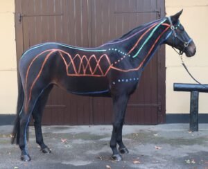
Research describing the Equine Myofascial Kinetic Lines (Harrison et al., 2015; Harrison and Elbrønd, 2018) has further highlighted the extensive continuity of fascial tissues throughout the horse’s body. These interconnected lines help explain why dysfunction observed in one anatomical region may sometimes be associated with compensatory changes elsewhere, reinforcing the importance of assessing the horse as a whole rather than focusing solely on the site of apparent discomfort (Harrison et al., 2015; Harrison and Elbrønd, 2018).
Although fascial adaptations are a normal response to training and mechanical loading, prolonged restriction of movement, injury or repetitive strain may alter tissue mobility and mechanical behaviour. However, the relationship between these changes and clinical signs such as pain or reduced performance remains an area of active scientific investigation, and many questions are still being explored (Schleip et al., 2012; Elbrønd and Schultz, 2021).
What is myofascial release?
Myofascial release is a physiotherapy technique that aims to influence the fascial system through the application of gentle, sustained or controlled mechanical pressure to soft tissues. The objective is not simply to “massage muscles”, but to address the interaction between muscles and their surrounding connective tissues in order to improve movement quality and tissue function (Cheatham et al., 2019).
Historically, myofascial release was thought to work primarily by physically stretching fascia or breaking down adhesions within connective tissues; but while these explanations remain common, current scientific understanding suggests that the mechanisms involved are likely to be far more complex.
Rather than mechanically “releasing” fascia alone, myofascial interventions are now thought to influence tissues through a combination of mechanical, neurological and cellular responses. Controlled mechanical loading may stimulate specialised cells within the fascia, known as fibroblasts, triggering processes involved in tissue adaptation and remodelling – a phenomenon known as “mechanotransduction”(Chaudhry et al., 2007; Meltzer et al., 2010). At the same time, manual therapy may influence the nervous system, altering muscle tone, movement patterns and pain perception (Schleip et al., 2012).
Importantly, these mechanisms continue to be investigated, and researchers acknowledge that the precise physiological effects of myofascial release have not yet been fully established. This does not mean the technique lacks value; rather, it highlights the need for continued high-quality research to better understand how, when and for whom these interventions are most effective (Cheatham et al., 2019; Seffrin et al., 2019).
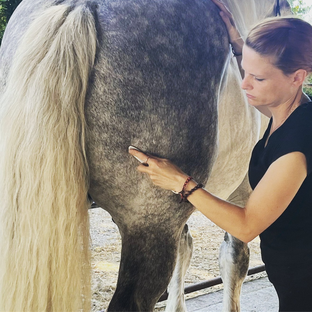
Today, myofascial release may be performed either manually, using the practitioner’s hands, or with specially designed instruments, often referred to as instrument-assisted soft tissue mobilisation (IASTM) or instrument-assisted myofascial release.
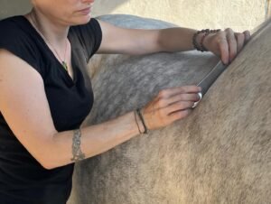
While the methods differ, both approaches apply controlled mechanical stimuli to soft tissues with the aim of improving movement and supporting normal musculoskeletal function. Current evidence does not suggest that one approach is universally superior; rather, the choice depends on the individual horse, the treatment objectives and the clinician’s assessment.
__________________________________________________________________________
REFERENCES
Adstrum, S., Hedley, G., Schleip, R., Stecco, C. and Yucesoy, C.A. (2017). Defining the fascial system. Journal of Bodywork and Movement Therapies, 21(1), pp.173–177.
Chaudhry, H., Schleip, R., Ji, Z., Bukiet, B., Maney, M. and Findley, T. (2007). Three-dimensional mathematical model for deformation of human fasciae in manual therapy. Journal of the American Osteopathic Association, 108(8), pp.379–390.
Cheatham, S.W., Lee, M., Cain, M. and Baker, R. (2019). The efficacy of instrument assisted soft tissue mobilization: A systematic review. Journal of the Canadian Chiropractic Association, 63(3), pp.200–211.
Elbrønd, V.S. and Schultz, R.M. (2015). Myofascial kinetic lines in the horse. Anatomia, Histologia, Embryologia, 44(6), pp.464–476.
Elbrønd, V.S. and Schultz, R.M. (2021). Fascia and the fascial system in the horse: A review. Journal of Equine Veterinary Science, 104, 103676.
Harrison, I., Elbrønd, V.S., Riis-Olesen, K. and Bartels, E.M. (2015). Multi-myofascial lines in the horse: Biomechanical, anatomical and physiological perspectives. Journal of Equine Veterinary Science, 35(9), pp.712–731.
Harrison, I. and Elbrønd, V.S. (2018). The equine myofascial kinetic lines: A comparative review. Journal of Equine Veterinary Science, 65, pp.90–98.
Meltzer, K.R., Cao, T.V., Schad, J.F., King, H., Stoll, S.T., Standley, P.R. and Findley, T.W. (2010). In vitro modeling of repetitive motion injury and myofascial release. Journal of Bodywork and Movement Therapies, 14(2), pp.162–171.
Schleip, R., Jäger, H. and Klingler, W. (2012). What is ‘fascia’? A review of different nomenclatures. Journal of Bodywork and Movement Therapies, 16(4), pp.496–502.
Schultz, R. M., Due, T. & Elbrønd, V. S. (2021). Equine Myofascial Kinetic Lines for Professionals – Equine Rehabilitation and Movement Anatomy, Myofascial Therapy, Biomechanics and Physiotherapy Guide for Veterinarians and Therapists. Kalmar, Sweden: Leanders Grafiska AB Kalmar.
Seffrin, C.B., Cattano, N.M., Reed, M.A. and Gardiner-Shires, A.M. (2019). Instrument-assisted soft tissue mobilization: A systematic review and effect-size analysis. Journal of Athletic Training, 54(7), pp.808–821.
Stecco, C., Schleip, R. and Huijing, P.A. (2018). Functional Atlas of the Human Fascial System. 2nd ed. Edinburgh: Elsevier.
Wilke, J., Krause, F., Vogt, L. and Banzer, W. (2018). What is evidence-based about myofascial chains? A systematic review. Archives of Physical Medicine and Rehabilitation, 99(3), pp.454–461.